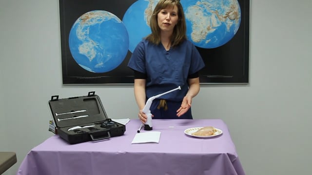
Thermal Ablation (Cold Probe) of Cervical Pre-Cancer Lesions
This Sexual and Reproductive Health and Rights (STARS) - Cervical Cancer Screening and Treatment skills module allows nurses, midwives, clinical officers, and medical officers to become confident and competent in performing thermal ablation of cervical pre-cancer lesions as part of cervical cancer screening and treatment procedures performed in primary health care facilities and mobile units in resource-constrained settings.
Learning Objectives[edit | edit source]
By the end of this module, learners will be able to perform thermal ablation following the correct steps for the Liger Medical HTU-110 Thermocoagulator™ which has a probe shaft and heating tip that is inserted and removed unheated ("cold").[1]
Materials and Equipment[edit | edit source]
Supplies[edit | edit source]
- Android Phone with Thermocoagulator Simulator Mobile App
- Pen
Gynecologic Simulator[edit | edit source]
The Gynecologic Simulator, Vaginal Canal with Augmented Feedback, and VIA Positive Cervix will be used in this skills module.
Thermocoagulator Simulator and Mobile App[edit | edit source]
The Liger Medical HTU-110 Thermocoagulator™ is designed with a probe shaft with heating tip that is inserted and removed unheated ("cold") to reduce the risk of burn injury to the patient.[1]
The Thermocoagulator Simulator with a cold probe is modelled on the Liger Medical HTU-110 Thermocoagulator™ and uses a Thermocoagulator Simulator Mobile App, cardboard device, and 16.0 mm and 19.0 mm diameter flat probes to simulate the thermal ablation procedure.
The Thermocoagulator Simulator also has an innovative augmented feedback feature to alert the user if the heated probe shaft or tip touches the vaginal sidewalls of the Gynecologic Simulator during the thermal ablation procedure.
Training Logbook[edit | edit source]
Please print out the Training Logbook below and write your name and date of training at the bottom of the Training Logbook page.
| # | Step | Check box if step was completed |
|---|---|---|
| 1 | Wash hands thoroughly and air dry them | |
| 2 | Put on gloves | |
| 3 | Position the light source so that the cervix can be visualized clearly | |
| 4 | On the VIA Positive Cervix in the Gynecologic Simulator, identify theː*
|
|
| 5 | Photograph the pre-treatment cervix on a cellphone* | |
| 6 | Select a probe of appropriate size to cover the cervix | |
| 7 | Apply the probe on the area of the cervix to be treated | |
| 8 | Ensure that the probe does not contact the vaginal wall during the treatment and cool down cycles* | |
| 9 | Check if the entire Transitional Zone has been treated | |
| 10 | If not, then repeat the procedure so as to treat the entire Transitional Zone including the lesion on the ectocervix (1–5 overlapping applications can be used) | |
| 11 | For repeated procedures, ensure that the probe does not contact the vaginal wall during the treatment and cool down cycles* | |
| 12 | Photograph the post-treatment cervix on a cellphone* | |
| 13 | Remove gloves | |
| 14 | Wash hands thoroughly with soap and water and air dry them | |
| 15 | Complete the documentation to record the treatment[4] in the space on the rightː*
|
*These checklist items must have been completed by the learner to pass this module.
Learner's Nameː
Evaluator's Nameː
Date of Trainingː
Thermal Ablation (Cold Probe) Step By Step[edit | edit source]
*The steps highlighted in bold and with an asterix (̈*) are considered as critical.[2][3]
- Check that instruments, supplies and light source are available and ready to use.[2][3]
- Check that the Thermocoagulator Simulator, Thermocoagulator Simulator Mobile App, Gynecologic Simulator and VIA Positive Cervix are ready for use.*
- Wash hands thoroughly with soap and water and air dry them.
- Put on gloves.
- Ask a colleague to observe you and fill out the Training Logbook checklist.
- Open the videocamera function on the smartphone camera.
- Turn on the flash.
- Move the smartphone light source so that the cervix can be visualized clearly.
On the VIA Positive Cervix, identify:*[2][3]
- Squamocolumnar Junction
- Limits of the lesion
- Transitional Zone and area to treat
- Press on the back button twice on the smartphone to return to the main screen
- Open the Thermocoagulator Simulator Mobile App and press on the "Take Pre-Treatment Photo" button which activates the smartphone's camera.
- Turn on the camera flash.
- Photograph the cervix pre-treatment.
- Press on the back button twice on the smartphone to return to the main screen of the Thermocoagulator Simulator Mobile App.
- Select a cold probe of appropriate size (16.0 mm or 19.0 mm)[1] to cover the cervix.
- Connect the probe to the Thermocoagulator Simulator.
- Cut a small length of small gauge wire.
- Loop and twist together the two ends of the small gauge wire around the aluminum foil covering the proximal probe shaft of the Thermocoagulator Simulator.
- Attach one alligator clip to the wire loop around the proximal probe shaft.
- Wind this alligator clip down the front of the Thermocoagulator Simulator and around the handle of the Thermocoagulator Simulator.
- Tape a section of the alligator clip that winds around the handle of the Thermocoagulator Simulator.
- Attach a second alligator clip to the free end of the alligator clip attached to the wire loop around the proximal probe shaft.
- In the main screen of the Thermocoagulator Simulator Mobile App, press on the "Run Simulation" button.
- Turn on the Thermocoagulator Simulator by pressing the ON/OFF button once.[1]
- Verify that the green light in the center of the ON/OFF button is on.
- The white illumination light on the back of the smartphone will also turn on.
- One blue light will flash showing the Thermocoagulator Simulator is ready for placement (tip is not heated yet).
- Apply the Thermocoagulator Simulator probe on the area of the cervix to be treated.*[2][3]
- Make sure that the probe is not touching the vaginal sidewalls.*
- Connect the alligator clip (attached to the exposed wire of the red insulated end of the buzzer) of the Augmented Feedback Circuit to the exposed aluminum foil of the Vaginal Canal.
- Connect the alligator clip from the Thermocoagulator Simulator to the other (unused) lead of a 9 V battery.
- While the probe is placed against the tissue needing treatment, press the ON/OFF button a second time to start the procedure.[1]
- Four blue timer lights will flash sequentially from left to right for a few seconds (~8 seconds) indicating that the probe tip is heating to 100°C.
- When the blue timer lights turn solid and a single audible beep is heard, the treatment cycle is running.
- The mobile app will produce a slight or faint popping noise to simulate the sounds that can be heard during thermal ablation.
- The blue timer lights turn off with an audible beep, one at a time, after each 1/4 of the procedure has finished (5 seconds).
- Make sure that the probe is not touching the vaginal sidewalls during the treatment cycle.* The buzzer will alarm if the heated probe contacts the vaginal sidewalls.
- When all four (4) blue timer lights are turned off a longer audible beep is heard, the unit is no longer applying heat (~20 seconds). It has commenced its cool down cycle (~10 seconds).[1]
- Make sure that the probe is not touching the vaginal sidewalls during the cool down cycle.* The buzzer will alarm if the heated probe contacts the vaginal sidewalls.
- Once the cool down cycle is complete, the white illumination light of the smartphone will turn off.
- Turn off the Thermocoagulator Simulator by pressing the ON/OFF button once.
- Verify that the green light in the center of the ON/OFF button is off.
- Press on the back button once on your smartphone to return to the main screen of the Thermocoagulator Simulator Mobile App.
- Disconnect the alligator clips from the aluminum foil of the Vaginal Canal with Augmented Feedback and the Thermocoagulator Simulator.
- Remove probe from the treatment area.
- Repeat Step 5 and check if the entire Transitional Zone has been treated.[2][3]
- If not, then repeat Steps 6-9 of the procedure so as to treat the entire Transitional Zone including the lesion on the ectocervix (1–5 overlapping applications can be used).
- Once the entire Transitional Zone has been treated, press on the back button on your smartphone to return to the main screen of the Thermocoagulator Simulator Mobile App.
- Press on the "Take Post-Treatment Photo" button in the Thermocoagulator Simulator Mobile App.
- Turn on the camera flash.
- Photograph the cervix post-treatment.
- Press on the back button twice on the smartphone to return to the main screen of the smartphone.
- Power off the smartphone to minimize battery usage.
- Dissemble the Thermocoagulator Simulator for simulated cleaning and disinfection in accordance with the manufacturer's instructions.[1]
- Remove gloves.[2][3]
- Wash hands thoroughly with soap and water and dry with clean, dry cloth or air dry.
- Complete the documentation to record the treatment.*[2][3]
- Draw a map of the cervix and the treated area for Step #15 in the Training Logbook using the nomenclature of the International Federation of Cervical Pathology and Colposcopy (2011 IFCPC nomenclature).[4]
- Draw two concentric circles.
- The outer circle represents the boundary of the cervix, and the inner circle represents the external os.
- Divide the circle into four quadrants with two perpendicular lines.
- Draw the location of the squamocolumnar junction with a dotted line.
- Draw the acetowhite lesions as a continuous line.
- Draw the treatment area as a shaded area, showing its position in relation to the quadrants and the external os.
Self assessment[edit | edit source]
- Fill out the Training Logbook - Thermal Ablation (Cold Probe) of Cervical Pre-Cancer Lesions to complete this module's self-assessment framework.
Resources[edit | edit source]
Sections of the STARS - Cervical Cancer Screening and Treatment module are copied or adapted from: Training of health staff in VIA, HPV detection test and cryotherapy - Trainees' handbook. New Delhi: World Health Organization, Regional Office for South-East Asia; 2017. Licence: CC-BY-NC-SA-3.0 IGO; and Training of health staff in VIA, HPV detection test and cryotherapy - Facilitators' guide. New Delhi: World Health Organization, Regional Office for South-East Asia; 2017. Licence: CC-BY-NC-SA-3.0 IGO. The World Health Organization (WHO) is not responsible for the content or accuracy of any translation. The original English edition shall be the binding and authentic edition.
- ↑ 1.0 1.1 1.2 1.3 1.4 1.5 1.6 HTU-IFU-002 TC Thermocoagulator Instructions for Use Rev B 04/2021 CN 0354 [product insert]. Liger Medical; 2021.
- ↑ 2.0 2.1 2.2 2.3 2.4 2.5 2.6 2.7 Training of health staff in VIA, HPV detection test and cryotherapy - Trainees' handbook. New Delhi: World Health Organization, Regional Office for South-East Asia; 2017. Licence: CC-BY-NC-SA-3.0 IGO. The World Health Organization (WHO) is not responsible for the content or accuracy of any translation. The original English edition shall be the binding and authentic edition.
- ↑ 3.0 3.1 3.2 3.3 3.4 3.5 3.6 3.7 Training of health staff in VIA, HPV detection test and cryotherapy - Facilitators' guide. New Delhi: World Health Organization, Regional Office for South-East Asia; 2017. Licence: CC-BY-NC-SA-3.0 IGO. The World Health Organization (WHO) is not responsible for the content or accuracy of any translation. The original English edition shall be the binding and authentic edition.
- ↑ 4.0 4.1 Basu P, Sankaranarayanan R (2017). Atlas of Colposcopy – Principles and Practice: IARC CancerBase No. 13 [Internet]. Lyon, France: International Agency for Research on Cancer. Available from: https://screening.iarc.fr/atlascolpodetail.php?Index=28&e=,0,1,2,3,8,10,15,19,30,31,43,46,47,60,61,68,73,83,88,89,93,96,102,105,111#0, accessed on October 10, 2021.


