Pediatric Distal Forearm Fractures/Self-Assessment Framework

This module allows traditional bone setters, pre-hospital providers, clinical officers, nurses, nurse practitioners, and medical officers to become confident and competent in performing point-of-care ultrasound diagnostic imaging to rule out the presence of a pediatric distal forearm fracture and distinguish between buckle (torus) fractures and cortical break fractures to make appropriate referrals as part of the management of closed pediatric (< 16 years of age) distal forearm fractures in regions without access to X-ray imaging and orthopedic specialist coverage.[1][2][3][4][5][6][7][8][9]
Self-Assessment Framework[edit | edit source]
Please print off this self-assessment framework before the simulation skills training, review it after the simulation skills training, and file and save a back-up copy of the completed and signed self-assessment framework and your ultrasound images for your training records.
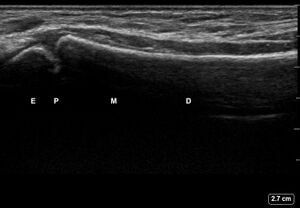
Review all the acquired images to determine if they meet all the quality standards outlined in the checklist below.
| # | Proper Technique for Image Acquisition and Interpretation | Ultrasound Image Meets Standard | Ultrasound Image Does Not Meet Standard |
|---|---|---|---|
| 1 |  |
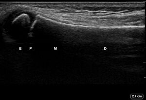 |
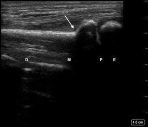 |
| 2 | 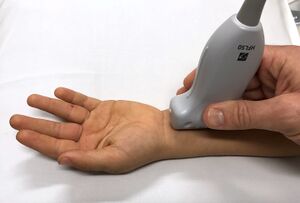 |
 |
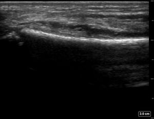 |
| 3 |  |
 |
 |
| 4 | Probe is set to the proper depth of 4 cm when evaluating the pronator quadratus muscle. |  |
 |
| 5 | All features are properly labelled. | 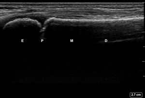 |
 |
Any image that does not meet the standard for all the quality assurance checklist items must be re-acquired in accordance with the techniques described below.
| Ultrasound Scanning Site | Please check off each ultrasound view that meets all 5 quality assurance checklist standards |
|---|---|
| Left Forearm of Healthy Adult |
View #1: Dorsal Radius View #2: Dorsal Ulna View #3: Lateral Ulna View #4: Lateral Radius View #5: Volar Radius View #6: Volar Ulna View of Pronator Quadratus |
| Right Forearm of Healthy Adult |
View #1: Dorsal Radius View #2: Dorsal Ulna View #3: Lateral Ulna View #4: Lateral Radius View #5: Volar Radius View #6: Volar Ulna View of Pronator Quadratus |
| Models #1 + #2 |
View #1: Dorsal Radius View #2: Dorsal Ulna View #3: Lateral Ulna View #4: Lateral Radius View #5: Volar Radius View #6: Volar Ulna |
| Models #3 + #4 |
View #1: Dorsal Radius View #2: Dorsal Ulna View #3: Lateral Ulna View #4: Lateral Radius View #5: Volar Radius View #6: Volar Ulna |
| Models #5 + #6 (Unblinded Training) |
View #1: Dorsal Radius View #2: Dorsal Ulna View #3: Lateral Ulna View #4: Lateral Radius View #5: Volar Radius View #6: Volar Ulna |
| Models #5 + #6 (Blinded Training) |
View #1: Dorsal Radius View #2: Dorsal Ulna View #3: Lateral Ulna View #4: Lateral Radius View #5: Volar Radius View #6: Volar Ulna |
| Models #7 + #8 |
View #1: Dorsal Radius View #2: Dorsal Ulna View #3: Lateral Ulna View #4: Lateral Radius View #5: Volar Radius View #6: Volar Ulna |
| Models #9 + #10 |
View #1: Dorsal Radius View #2: Dorsal Ulna View #3: Lateral Ulna View #4: Lateral Radius View #5: Volar Radius View #6: Volar Ulna |
All images must meet all the quality assurance standards checklist in order to pass this skills training module.
Learner's Nameː
Learner's Signature:
Assistant's Nameː
Assistant's Signature:
Date of Trainingː
Training Module Certificate of Completion[edit | edit source]
Once all the ultrasound images have been reviewed and confirmed they all meet standards in the quality assurance checklist, then:
- Click the button above.
- Type in your name, download and print out a certificate of completion for this training module.
- Photograph your certificate on your cellphone as a backup and file the printed certificate in your training records.
Acknowledgements[edit | edit source]
This work is funded by a grant from the Intuitive Foundation. Any research, findings, conclusions, or recommendations expressed in this work are those of the author(s), and not of the Intuitive Foundation.
References[edit | edit source]
- ↑ Onyemaechi NO, Itanyi IU, Ossai PO, Ezeanolue EE. Can traditional bonesetters become trained technicians? Feasibility study among a cohort of Nigerian traditional bonesetters. Hum Resour Health. 2020 Mar 20;18(1):24. doi: 10.1186/s12960-020-00468-w. PMID: 32197617; PMCID: PMC7085192.
- ↑ Heiner JD, McArthur TJ. The ultrasound identification of simulated long bone fractures by prehospital providers. Wilderness Environ Med. 2010 Jun;21(2):137-40. doi: 10.1016/j.wem.2009.12.028. Epub 2009 Dec 22. PMID: 20591377.
- ↑ Heiner JD, Baker BL, McArthur TJ. The ultrasound detection of simulated long bone fractures by U.S. Army Special Forces Medics. J Spec Oper Med. 2010 Spring;10(2):7-10. PMID: 20936597.
- ↑ Heiner JD, Proffitt AM, McArthur TJ. The ability of emergency nurses to detect simulated long bone fractures with portable ultrasound. Int Emerg Nurs. 2011 Jul;19(3):120-4. doi: 10.1016/j.ienj.2010.08.004. Epub 2010 Sep 25. PMID: 21665155.
- ↑ Snelling PJ, Jones P, Keijzers G, Bade D, Herd DW, Ware RS. Nurse practitioner administered point-of-care ultrasound compared with X-ray for children with clinically non-angulated distal forearm fractures in the ED: a diagnostic study. Emerg Med J. 2021 Feb;38(2):139-145. doi: 10.1136/emermed-2020-209689. Epub 2020 Sep 8. PMID: 32900856.
- ↑ Snelling PJ, Jones P, Moore M, Gimpel P, Rogers R, Liew K, Ware RS, Keijzers G. Describing the learning curve of novices for the diagnosis of paediatric distal forearm fractures using point-of-care ultrasound. Australas J Ultrasound Med. 2022 Mar 7;25(2):66-73. doi: 10.1002/ajum.12291. PMID: 35722050; PMCID: PMC9201201.
- ↑ Heiner JD, McArthur TJ. A simulation model for the ultrasound diagnosis of long-bone fractures. Simul Healthc. 2009 Winter;4(4):228-31. doi: 10.1097/SIH.0b013e3181b1a8d0. PMID: 19915442.
- ↑ Snelling PJ, Keijzers G, Byrnes J, Bade D, George S, Moore M, Jones P, Davison M, Roan R, Ware RS. Bedside Ultrasound Conducted in Kids with distal upper Limb fractures in the Emergency Department (BUCKLED): a protocol for an open-label non-inferiority diagnostic randomised controlled trial. Trials. 2021 Apr 14;22(1):282. doi: 10.1186/s13063-021-05239-z. PMID: 33853650; PMCID: PMC8048294.
- ↑ Snelling PJ. A low-cost ultrasound model for simulation of paediatric distal forearm fractures. Australas J Ultrasound Med. 2018 Feb 25;21(2):70-74. doi: 10.1002/ajum.12083. PMID: 34760505; PMCID: PMC8409885.
