3D Printed Adult Male Tibial Bone Models/3D Print Sample Model

These 3D printed models accurately simulate bone length and diameter, external contour, cross-sectional shape, bicortical anatomy, cortical hardness, cancellous bone porosity, and microstructure, and far cortex thickness for adult, non-obese males at left tibial shaft fracture pin drilling sites for modular external fixation.[1][2][3][4][5][6][7][8][9][10][11][12] These models feature a semi-engraved model number, gender symbol, and drilling direction arrow on the base of each model to assist with model identification and proper orientation. Each model has a vise attachment to allow the user to secure the model inside a standard vise clamp to maximize safety during simulation training.
3D Print Sample Models[edit | edit source]
For optimal print results, use brand new white PLA filament fresh out of the sealed package to 3D print each sample model.
- 3D print one sample Adult Male Tibial Bone Model #1 and #2.
- Print the quality assurance checklist below for each 3D printed sample model by clicking on the "Download PDF" button in the upper righthand corner of this screen and selecting "Layout: Landscape" option for printing.
- Fill out the quality assurance checklist to verify that each sample model was properly printed.
- Write the date of inspection and print and sign your name at the bottom of the checklist.
- File and save a back-up copy of the completed and signed checklist for your production and inspection records.
| # | Action | Meets Standard | Does Not Meet Standard | Does Not Meet Standard | Check most appropriate response |
|---|---|---|---|---|---|
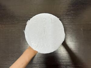
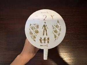
| 1 | Inspect the base. | 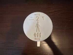 |
 |
 |
🔲 Meets Standard 🔲 Does Not Meet Standard |
| 2 | Inspect the top. | 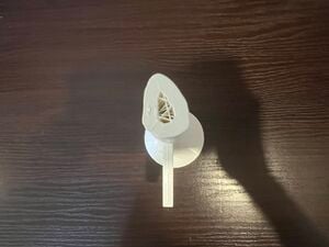 |
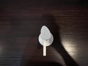 |
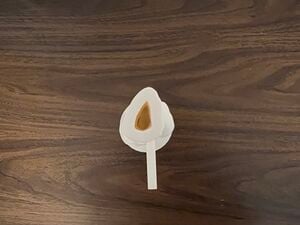 |
🔲 Meets Standard 🔲 Does Not Meet Standard |
| 3 | Inspect sides. | 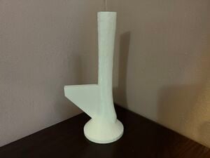 |
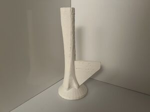 |
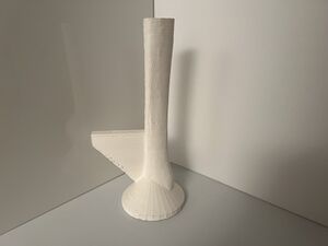 |
🔲 Meets Standard 🔲 Does Not Meet Standard |
| 4 | Inspect the vise attachment. | 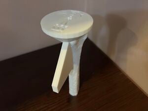 |
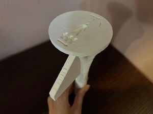 |
 |
🔲 Meets Standard 🔲 Does Not Meet Standard |
Each model must meet standards for all the quality assurance checklist items in order to be acceptable for the Tibial Fracture Fixation skills modules.
Adult Male Tibial Bone Model # (circle one): 1 or 2
Quality Assurance Checklist Completion Date (Day-Month-Year):
Inspected By (First Name and Last Name):
Signature:
Acknowledgements[edit | edit source]
This work is funded by a grant from the Intuitive Foundation. Any research, findings, conclusions, or recommendations expressed in this work are those of the author(s), and not of the Intuitive Foundation.
References[edit | edit source]
- ↑ Ugochukwu EG, Ugbem LP, Ijomone OM, Ebi OT. Estimation of Maximum Tibia Length from its Measured Anthropometric Parameters in a Nigerian Population. J Forensic Sci Med [serial online] 2016 [cited 2021 Jun 27];2:222-8. Available from: https://www.jfsmonline.com/text.asp?2016/2/4/222/197928.
- ↑ U.S. Department of Health and Human Services — National Institutes of Health. Human tibia and fibula. [Internet]. Bethesda, (MD): NIH 3D Print Exchange; 2014 May 29 [cited 2021 Aug 17]. Available from: https://3dprint.nih.gov/discover/3DPX-000169.
- ↑ Gosman JH, Hubbell ZR, Shaw CN, Ryan TM. Development of cortical bone geometry in the human femoral and tibial diaphysis. Anat Rec (Hoboken). 2013 May;296(5):774-87. doi: 10.1002/ar.22688. Epub 2013 Mar 27. PMID: 23533061.
- ↑ www.prusa3d.com/file/370474/technical-data-sheet.pdf
- ↑ Ultimaker. Ultimaker PLA Technical Data Sheet [Internet]. Ultimaker Support. [cited 2021 July 29]. Available from: https://support.ultimaker.com/hc/en-us/articles/360011962720-UltimakerPLA-TDS.
- ↑ https://support.ultimaker.com/hc/en-us/articles/360011962720-Ultimaker-PLA-TDS
- ↑ Vian, Wei Dai and Denton, Nancy L., "Hardness Comparison of Polymer Specimens Produced with Different Processes" (2018). ASEE IL-IN Section Conference. 3. https://docs.lib.purdue.edu/aseeil-insectionconference/2018/tech/3.
- ↑ Society For Biomaterials 30th Annual Meeting Transactions, page 332. Femoral Cortical Wall Thickness And Hardness Evaluation. K. Calvert, L.A. Kirkpatrick, D.M. Blakemore, T.S. Johnson. Zimmer, Inc., Warsaw, IN.
- ↑ Meyers, M. A.; Chen, P.-Y. (2014). Biological Materials Science. Cambridge: Cambridge University Press. ISBN 978-1-107-01045-1.
- ↑ Forrest AM, Johnson AE, inventors; Pacific Research Laboratories, Inc., assignee. Artificial bones and methods of making same. United States patent 8,210,852 B2. Date issued 2012 Jul 3.
- ↑ National Institutes of Health Osteoporosis and Related Bone Diseases National Resource Center. What is Bone? [Internet]. Bethesda (MD): The National Institutes of Health (NIH); 2018. [Cited 2021 Aug 17]. Available from: https://www.bones.nih.gov/health-info/bone/bone-health/what-is-bone.
- ↑ Maeda K, Mochizuki T, Kobayashi K, Tanifuji O, Someya K, Hokari S, Katsumi R, Morise Y, Koga H, Sakamoto M, Koga Y, Kawashima H. Cortical thickness of the tibial diaphysis reveals age- and sex-related characteristics between non-obese healthy young and elderly subjects depending on the tibial regions. J Exp Orthop. 2020 Oct 6;7(1):78. doi: 10.1186/s40634-020-00297-9. PMID: 33025285; PMCID: PMC7538524.
